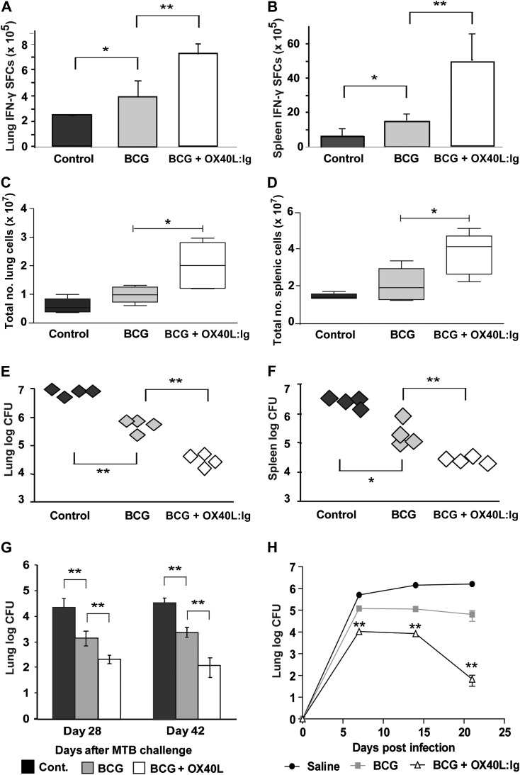
Figure 3.
The role of OX40/OX40L in immunity to BCG and Mycobacterium tuberculosis (M. tuberculosis) challenge. Five mice per group were inoculated subcutaneously with phosphate-buffered saline (cont.), BCG plus control immunoglobulin (Ig) (BCG), or BCG + OX40L:Ig and challenged with 5 × 10⁵ M. tuberculosis (strain R37rv) intravenously or 50–100 bacilli by aerosol. In mice intravenously challenged 42 days after vaccination, interferon (IFN)–γ spot-forming colonies (SFCs) from purified protein derivative –stimulated lung cells (A) or splenocytes (B) was enumerated by enzyme-linked immunosorbent spot assay (mean + SD of 5 mice per group) 14 days after challenge. In the same mice, total numbers of viable cells (C and D) and colony-forming units (CFUs) (E and F) in the lung (C and E) and spleen (D and F) 14 days after M. tuberculosis challenge were determined. In aerosol-challenged mice (G and H), the number of M. tuberculosis CFUs was determined by diluting homogenates of lung in mice challenged with M. tuberculosis 28 days (G) or 180 days (H) after BCG vaccination. All graphs show results for 4–5 mice per group and are representative of 2–3 independent experiments. *P < .05, **P < .01 by Mann–Whitney test (in H, significance is presented for BCG vs BCG + OX40L:Ig).

