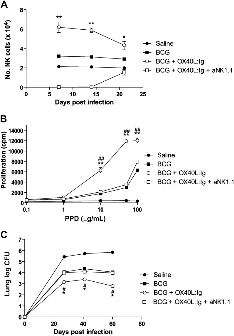
Figure 4.
A critical role for natural killer (NK) cells in OX40L:Ig-mediated protection from Mycobacterium tuberculosis (M. tuberculosis). C57BL/6 mice were inoculated subcutaneously with saline (closed circle) or 2 × 10⁷ BCG coadministered with intraperitoneal Control Ig (closed square), OX40L:Ig (open circle) or OX40L:Ig/anti-NK1.1 (open square). NK cells were examined by flow cytometry (CD4−CD8−DX5+) in the spleen at indicated time points after BCG vaccination (A). At 14 days after BCG vaccination, proliferative responses of spleen cells from each group were adjudged by H-3-thymidine incorporation in response to purified protein derivative (PPD) (B). (C) Groups of mice were challenged with intravenous injection 6 weeks after BCG administration and the number of colony-forming units (CFU) of M. tuberculosis was determined by diluting homogenates of lung at 25, 40, and 60 days after challenge. All graphs show results for 5–6 mice per group and are representative of 2–3 independent experiments. *P < .05, **P < .01 by Mann–Whitney U test for BCG vs BCG + OX40L:Ig. #P < .05, ##P < .01 by Mann–Whitney U test for BCG + OX40L:Ig vs BCG + OX40L:Ig + anti-NK1.1.

