Figure 4.
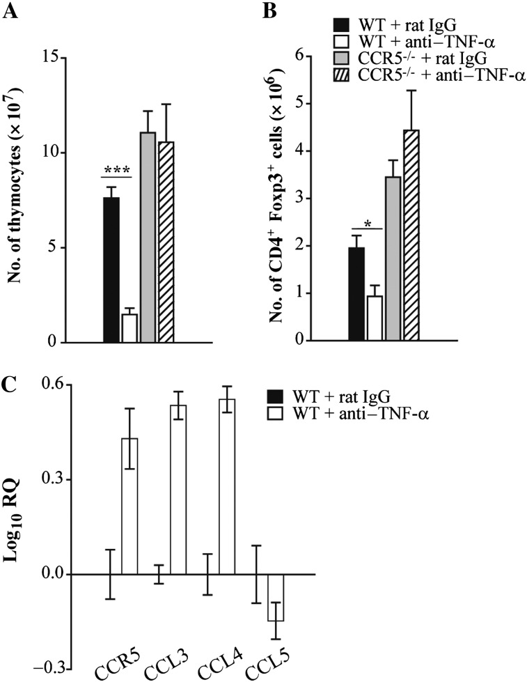
Tumor necrosis factor α (TNF-α) antagonism alters thymic cellularity in wild-type (WT) and, to a lesser extent, CCR5–/– mice during infection. Wild-type and CCR5–/– mice were given a monoclonal antibody to TNF-α or rat immunoglobulin G (IgG) and intranasally infected with 2 × 10⁶ yeasts. At day 7 postinfection, thymi were isolated from mice to determine the total number of thymocytes (A) and number of CD4+ Foxp3+ cells (B). RNA was extracted from lung leukocytes at day 7 postinfection and CCR5, CCL3, CCL4, and CCL5 expression was measured by quantitative real-time polymerase chain reaction. Hypoxanthine-guanine phosphoribosyltransferase was used as an endogenous control, and values represent log increase compared with IgG control lungs (C). Data represent the mean ± SEM (n = 6–12) from 2–3 experiments. **P < .01; ***P < .001.

