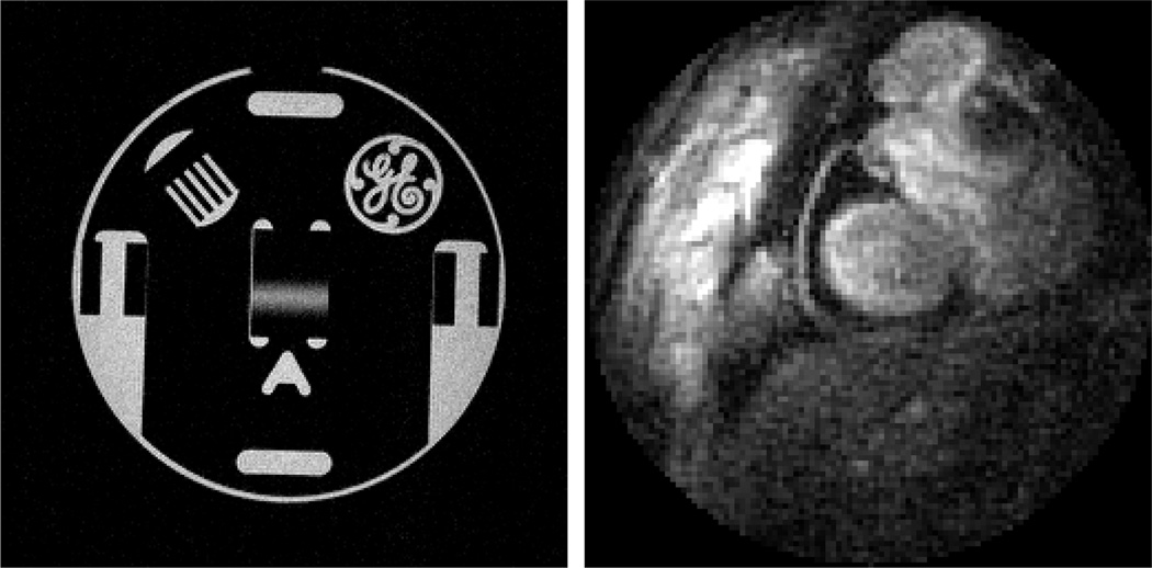
Fig. 13.
Left: MR image of 12cm GE resolution phantom, acquired using Medusa controlling a GE Signa Excite 1.5T scanner. Right: A frame capture of real-time cardiac imaging at 50 frames per second performed using a Medusa Console and RT-Hawk software suite on the 1.5T GE magnet. Acquisition is a 3072-pt spiral with 4× interleave at 20ms TR.

