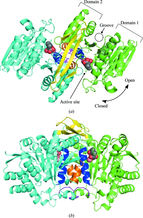
Figure 3.
Overall structure of the SoIPMDH–IPM complex. (a) Cartoon representation of the overall structure of the SoIPMDH dimer at 0.1 MPa (green, subunit A; cyan, subunit B; blue, helix g; orange, helix h; magenta, FG loop; yellow, arm-like region). (b) Image rotated by 90° compared with (a). The IPM substrate molecules and the calcium cations located in the active site of each subunit are represented as spheres.

