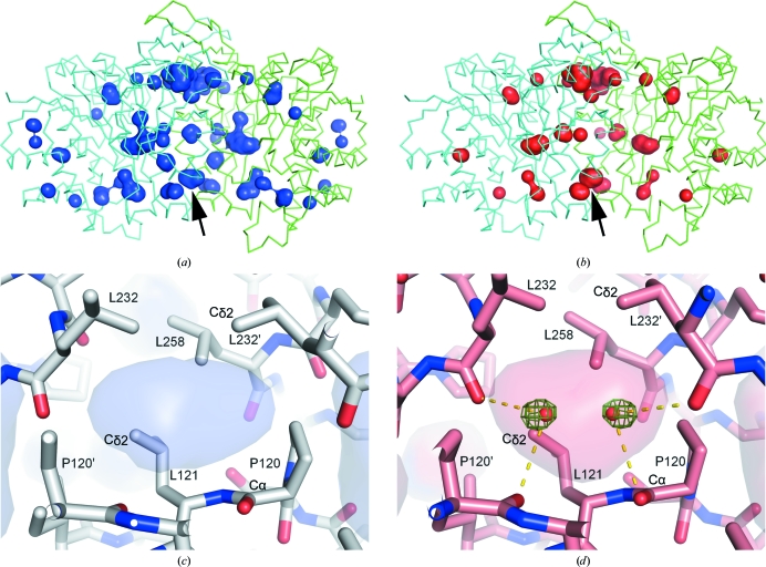
Figure 6.
Internal cavities of the SoIPMDH dimer and observed water penetration. (a) and (b) show internal cavities of the SoIPMDH dimer as surface representations at 0.1 and 580 MPa, respectively. The cavity for which the volume increases with pressure is indicated by an arrow. (c) and (d) are magnified views around the cavity at 0.1 and 580 MPa, respectively. A difference electron-density map is shown as a green mesh contoured at 4.0σ. No positive peaks are observed in (c) at 0.1 MPa. The transparent surfaces in (c) and (d) represent the cavities; hydrogen bonds are also shown in (d).

