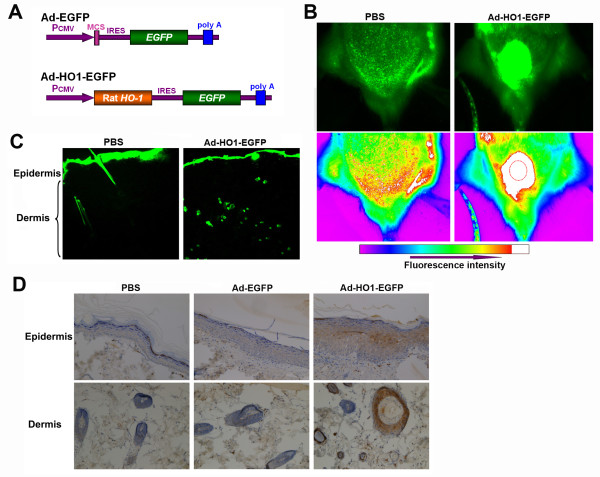
Figure 1.
The distribution and expression of Ad-HO1-EGFP in rat skin. (A) Schematic diagram of the recombinant Ad-HO1-EGFP vector and the control vector (Ad-EGFP). (B) In vivo imaging of EGFP expression in SD rats. Rats were injected subcutaneously with 5 × 10⁹ genomic copies of Ad-HO1-EGFP or equivalent volume of PBS. Three days after injection, the expression of EGFP was visualized. The dotted circle indicates the injection region. (C) The expression of EGFP was imaged from frozen-cut sections and observed under a fluorescent microscope (× 40). (D) Immunohistochemistry analysis of HO-1 expression in rat epidermis and dermis (× 200).

