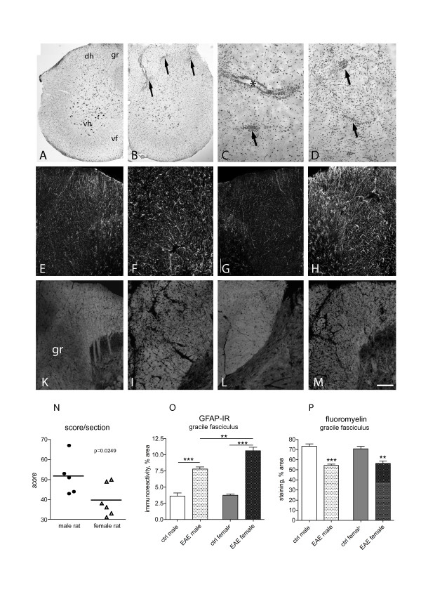
Figure 2.
Histopathology of the spinal cord in control and EAE animals was performed by E&E (A-D), GFAP-immunostaining for astroglial reaction (E-H) and FluoroMyelin staining for white tractd and myelin sheaths (K-M and related inserts). Micrograph in A illustrates a spinal cord hemi-sections in control, male rat; micrograph in B a spinal cord hemi-sections showing the extensive inflammatory infiltrates in EAE, as detailed in high power micrographs in C and D. Arrows indicate the intraparenchimal infiltrates asterisk in C a perivascular infiltrate. Image analysis indicates that there is a difference in the extension of inflammation infiltrate in female vs male EAE animals (N, Student's t test, p = 0.00249). Micrographs from E to H illustrate the astroglial reaction in EAE animals (F: female; H: male) with control animals (E: female; G: male). Image analysis indicates that astroglial reaction is stronger in female than in male EAE animals (O, one way ANOVA and Tukey multiple comparison test, **p < 0.01; ***p < 0.001). Micrographs from K to M illustrate white matter in EAE (I: female; M: male) compared with control animals (K: female; L: male). The color inserts refer to high power magnification, to shown the myelin sheath morphology. The original image has been processed using a deconvolution procedure. Image analysis indicates that demyelination is not different in female and male EAE animals (one way ANOVA and Tukey multiple comparison test, **p < 0.01; ***p < 0.001). Graph bars: M, 100 μm; color insert 10 μm.

