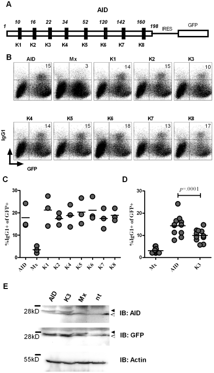
Figure 1. Effect of lysine to arginine mutations in the AID protein on CSR activity.
(A) Schematic representation of AID highlighting the position of the lysine (K) residues. For CSR assays, untagged constructs were expressed bicistronically together with GFP separated by an internal ribosome entry site (IRES). (B) Representative FACS profile for CSR activity of individual Lys to Arg mutants. B cells from AID deficient mice were retrovirally transfected with the respective lysine AID mutant (Mx: empty vector) and CSR to IgG1 was determined upon stimulation of cells with LPS and IL-4. GFP expression of infected cells was driven from an IRES element located 3′ of the stop codon of the cloned AID mutant. The percentage in the upper right quadrants indicate the percentage of IgG1+ cells among the GFP+ (i.e. retrovirally infected) population. (C) Results from three independent experiments of a screen of lysine mutants showing the percentage of IgG1+ cells among the GFP+ population. (D) Results from 11 independent experiments comparing the CSR activity of AID and K3. (E) Western Blot on lysates from B cells retrovirally transfected with the respective constructs. Black arrowheads indicate specific band. White arrowheads indicate nonspecific band. (IB: immune blot).

