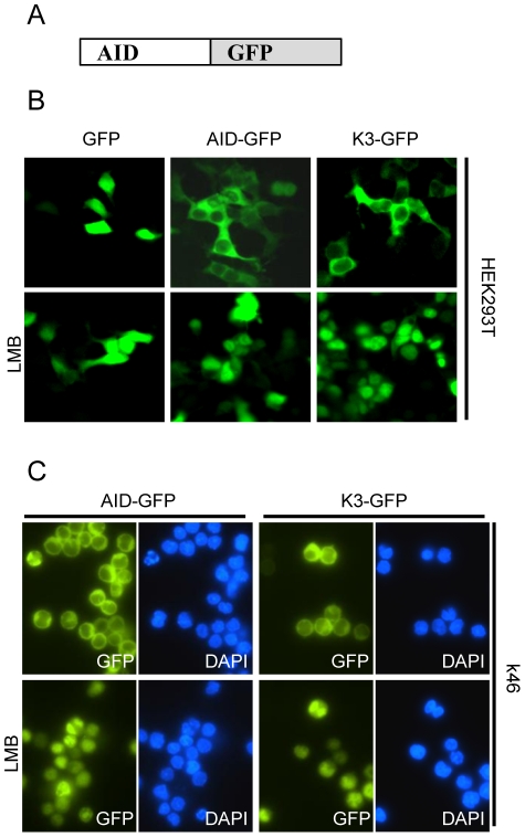
Figure 3. Subcellular localization of K3 is not different from wildtype AID.
(A) Schematic representation of AID-GFP fusion constructs used for localization assays. GFP is fused to the C-terminus of AID or mutant K3. (B) HEK293T cells were transiently transfected with the respective GFP-fusion constructs. Two days post transfection, cells were treated as indicated with Leptomycin B (LMB) for 3 hours. Cells were fixed and localization of the respective GFP fusion proteins visualized using fluorescence microscopy. (C) The k46 B cell line stably expressing AID-GFP or K3-GFP fusion proteins were treated with or without Leptomycin B (LMB) for 3 hours and localization of GFP fusion constructs was determined using fluorescence microscopy.

