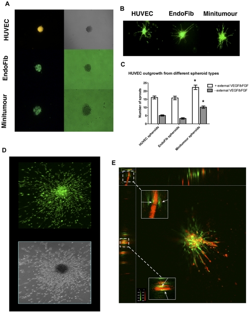
Figure 1. Characterization of the Minitumour spheroid model.
A - Fluorescent (left) and phase contrast (right) images of HUVEC, EndoFib and Minitumour spheroids before incubation in the collagen gel; endothelial cells pre-dyed with a CMFDA Green CellTracker dye are seen in each different spheroid type. B – Representative fluorescent images of spheroids after 48 h incubation in collagen gels, in the presence of complete medium, showing pre-dyed endothelial cells organized into pre-capillary sprouts. C – Quantification of endothelial sprout length from different spheroids show that MDA-MB-231 cells stimulate sprout formation even in the absence of exogenous growth factors VEGF and bFGF. D – Confocal (upper) and phase contrast (lower) images of MDA-MB231 cells pre-dyed with the green CellTracker dye in the Minitumour spheroid after 48 h incubation in complete medium. E - A 3D reconstruction of a Minitumour spheroid where the HUVECs have been dyed with a CMRA Orange CellTracker dye and the fibroblasts with a CMFDA Green Cell Tracker side panels show optical x and y sections of sprouts showing the deposition of HUVECs and Fibroblasts relative to sprout formation.

