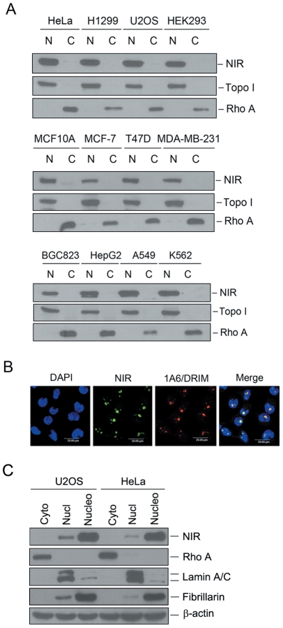
Figure 1. NIR is expressed in different human cell lines and mainly localized to the nucleolus.
A. Cytosolic and nuclear extracts were fractionized from multiple cancer cell lines as indicated at the top of blot. Same amount of protein was separated on SDS-PAGE, and transferred onto PVDF membranes. Blots were probed with anti-NIR. Fractionation was controlled by using nuclear marker protein topoisomerase I (Topo I) and a cytosolic marker protein RhoA. N represents nuclear extract and C represents cytoplasmic extract. B. Indirect immunofluorescence was performed with anti-NIR polyclonal antibody. NIR specific signal was recognized with FITC-conjugated goat anti-rabbit IgG. As a nucleolar protein, 1A6/DRIM was detected with anti-1A6/DRIM monoclonal antibody. 1A6/DRIM specific signal was recognized with TRITC-conjugated goat anti-mouse IgG. Nucleus was stained with DAPI. The image was obtained with confocal microscopy. C. Cytoplasmic, nucleoplasmic and nucleolar lysates were fractionized from U2OS and HeLa cells respectively. Same amount of protein from the above lysates was separated on SDS-PAGE, and transferred onto PVDF membranes. Blots were probed with anti-NIR. Rho A, lamin A/C and fibrillarin were used as controls for cytoplasmic, nucleoplasmic and nucleolar fractions respectively. Beta-actin was used as a loading control.

