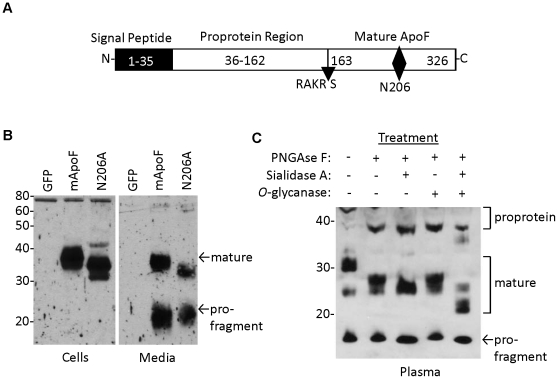
Figure 2. Glycosylation, processing and secretion of the mouse apo F protein.
A. Schematic of the mouse apo F precursor protein depicting the predicted boundaries of the signal peptide, proprotein region, and furin cleavage site (RAKR/S). The N-linked glycosylation site at N206 is shown with a diamond. B. Western blot for mouse apo F in cells (left) and media (right), from HEK293 cells transiently transfected with either green fluorescent protein (GFP), wild type mouse ApoF (mApoF), or mouse apo F with asparagine 206 mutated to alanine (N206A). C. Western blot of apo F in one microliter of plasma from mice overexpressing mApoF from a liver-specific AAV vector. Plasma was denatured with heat and then subjected with deglycosylation by PNGase F, Sialidase A, and O-glycanase. The mature and pro-fragment portions of the apo F protein are shown with arrows.

