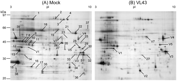
Figure 1. Typical 2-D gels showing Arabidopsis thaliana apoplastic leaf proteins after mock (A) or Verticillium longisporum .
(VL43) infection (B). Proteins were extracted 25 dpi and 80 µg were loaded for separation. Gels were stained with silver nitrate. Numbers in the gels indicate protein spots analyzed by ESI-LC/MS from preparative Coomassie-stained gels.

