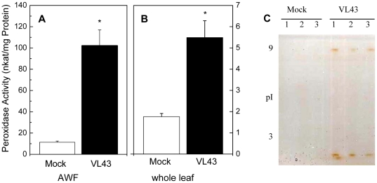
Figure 3. Peroxidase activity in apoplastic (A) and whole leaf extracts (B) and peroxidase activity staining in AWF (C).
Data are shown for mock (white columns) and Verticillium longisporum (VL43) infected Arabidopsis plants (black columns) 25 days post infection. Data indicate means (n = 6, ±SE). *indicate significant differences between mock- and VL-infected plants. For protein separation each lane of an isoelectric focusing gel (pH 3 to 9) was loaded with 3.5 µg AWF protein. Each of the three lanes of mock and VL43-infected plants corresponds to an independent biological replicate of AWF. After isoelectric focusing peroxidase activities were visualized by incubation in guaiacol/H2O2 solution.

