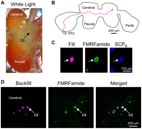
Figure 2. C2 characteristics in Tritonia diomedea.
A. C2Tri could be identified visually due to its characteristic white soma near the origin of cerebral nerve 1 (CeN1, white arrows). An asterisk (*) labels the statocyst. Only one cerebral-pleural ganglion is shown. B. Filling the C2Tri soma with Neurobiotin revealed a contralateral axon projection through the anterior cerebral-pedal commissure (not shown) and into the pedal commissure (PP2). The example is a representative image in which the outline of the brain and the axon projection were traced for ease of viewing. C. C2Tri (arrow) was filled with biocytin (left). It was immunoreactive for both FMRFamide (middle) and SCPB (right) as shown. D. Backfilling the pedal commissure with biocytin in Tritonia labeled 3–4 neurons near C2Tri (left). Only the cerebral-pleural ganglion contralateral to the backfilled nerve is shown. FMRFamide-like immunohistochemistry labeled C2Tri (middle). Combining FMRFamide-like immunoreactivity with the backfill revealed just one neuron, C2Tri, (right).

