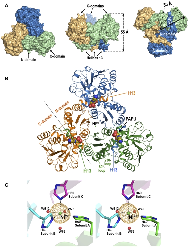
Figure 4. Structure of PTC trimer.
(A) Surface representations of the trimer along the threefold axis from the concave or convex faces (left and right, respectively) or with the threefold axis vertical and the concave face up (center). Each protomer is in a different color. (B) Cartoon representation of the PTC trimer viewed along its threefold axis with its concave face close to the viewer. PAPU is shown in space-filling representation. Some elements are labeled, and in one protomer the boundary between the N- and C-domains is signaled with a broken line. (C) Stereoview representation of the electron density omit map for the Ni ion (2.5σ) in one trimer of PTC-PALO, showing the coordinated histidine and water (marked W75, W76 and W512) molecules.

