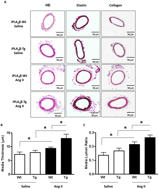
Figure 2. Exacerbated mesenteric artery remodeling in iPLA2β-Tg mice in response to Ang II infusion.
Vascular remodeling was analyzed in embedded sections of the 2nd order branch of mesenteric arteries from the iPLA2β-Wt and iPLA2β-Tg mice infused with saline or Ang II (500 ng/kg/min, 14 days). Representative images of vessel sections stained with HE, elastin or collagen (A). The media thickness (B) and media∶lumen ratio (C) were quantified. n = 8, *: p<0.05, **: p<0.01, ***: p<0.001 by one-way ANOVA.

