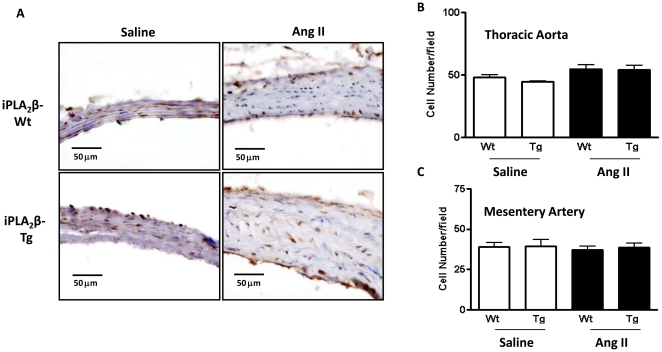
Figure 4. Lack of difference in cell proliferation between iPLA2β-Tg and iPLA2β-Wt mice by Ang II infusion.
Mice were infused with saline or Ang II (500 ng/kg/min, 14 days) and thoracic aortas were isolated and sectioned. (A) are representative images of the sections stained with anti-PCNA antibody. (B) and (C) are summaries of the cell number analysis using HE stained sections. n = 8.

