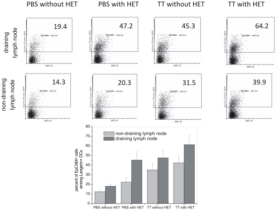
Figure 8. Migration of EpCAM+ epidermal DCs to the lymph node.
Mice were immunized by TT, with and without HET. Unimmunized (PBS without HET) animals served as control. Cell suspension from draining and non-draining lymph nodes were prepared after 24 h and stained with Langerin (CD207)-PE and EpCAM-Alexa Flour 488 antibodies. Stained cells were analyzed by flow cytometry, Langerin+ cells were gated and the expression of EpCAM was analyzed. Number on the selected area indicates percent of EpCAM+ cells among Langerin+ cells in total lymph node cells. The bar graph shows average percentages of three experiments.

