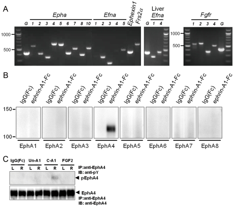
Figure 1. Expression of EphA–related signaling molecules in subventricular zone cells.
(A) RT-PCR for Ephas, Efnas, Fgfrs, Ephexin1, and Frs2α. G: Glyceraldehyde 6-phosphate dehydrogenase. For Efna1 and Efna4, liver RNA was also used as control. (B) Detection of EphA receptors that binds to ephrin-A1-Fc in rat brain lysate. Rat brain subventricular cell lysate was incubated with ephrin-A1-Fc, precipitated by protein A-agarose, and immunoblotted with the antibodies for EphAs. (C) EphA4 phosphorylation in tissue surrounding the lateral ventricle. In rats with lesioned unilaterally in the nigrostriatal dopaminergic pathway, subventricular tissue was collected 18 hours after single injection of clustered IgG(Fc) (3 µg), unclustered ephrin-A1-Fc (Un-A1) (3 µg), clustered ephrin-A1-Fc (C-A1) (3 µg), or FGF2 (100 ng). The tissue lysate (450 µg protein) was immunoprecipitated with anti-EphA4 antibody followed by immunoblotting with anti-phosphotyrosine (pY) antibody and anti-EphA4 antibody.

