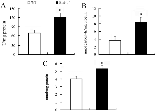
Figure 1. Brain oxidative stress in 4-week-old Bmi-1−/− mice.
Brain tissues from Bmi-1−/− mice showed higher levels of hydroxyl radical (120.82±14.72 vs. 69.41±10.04 U/mg protein; A), protein carbonyl (8.37±1.41 vs. 3.86±1.09 nmol/mg protein; B) and malondialdehyde (5.34±0.45 vs. 4.03±0.41 nmol/mg protein; C) than those from WT controls. Five mice per genotype and 3 independent experiments for each homogenized brain sample. Data are expressed as mean ± SEM. *P<0.05 vs. WT mice.

