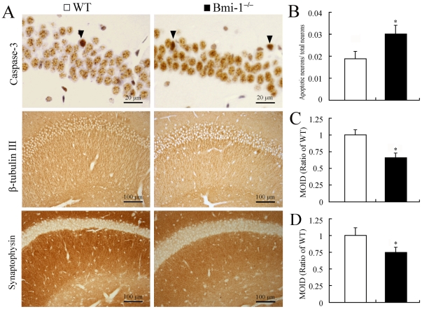
Figure 2. Degeneration of neuronal elements in the hippocampus of 4-week-old Bmi-1−/− mice.
(A) Immunostaining for caspase-3, β-tubulin III and synaptophysin. Arrowheads showing apoptotic pyramidal neurons which contained dense caspase-3 immunostaining. Immunoreactivities of β-tubulin III and synaptophysin were decreased in the Bmi-1 null hippocampus. (B–D) There were a higher ratio of apoptotic neurons to total neurons (0.031±0.004 vs. 0.019±0.003; B) and lower mean integrated optical densities (MIOD) of immunostainings for β-tubulin III (0.66±0.07 vs. 1±0.08; C) and synaptophysin (0.75±0.08 vs. 1±0.12; D) in the hippocampus of Bmi-1−/− mice compared with WT controls. Five mice per genotype and 3 sections per mouse. Data are expressed as mean ± SEM. *P<0.05 vs. WT mice.

