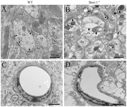
Figure 3. Ultrastructural alterations of the area CA1 stratum radiatum in 4-week-old Bmi-1−/− mice.
(A) A representative electron micrograph showing axonal (Ax)-dendrite (De) synapses (arrowheads) in WT mice. (B) Ultrastructural architecture of synaptic areas in Bmi-1−/− mice. The density of synapses decreased, and the residual synapses underwent degeneration (arrowheads). Many aberrant mitochondria (arrows) exhibited “zebra-like”, swollen or vacuolar profile. In addition, the density of microtubules also decreased in the dendritic cytoplasm as compared with that in the WT littermates. (C) A normal brain capillary from WT mice. Note that the capillary wall was surrounded by flattened endfeet of astrocytes (stars). (D) Abnormal architecture of the brain capillary from Bmi-1−/− mice. The capillary lumen was narrowed and obstructed accompanied with the swollen astrocyte endfeet (stars).

