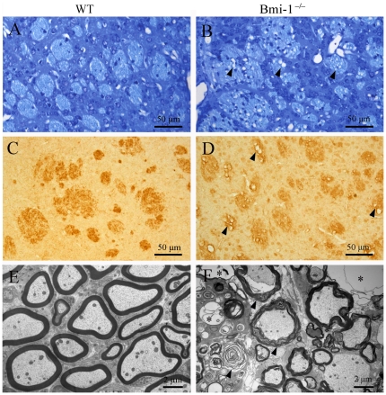
Figure 4. Demyelination in the striatum of 4-week-old Bmi-1−/− mice.
(A–D) Toluidine blue staining (A–B) and MBP immunohistochemistry (C–D). Many fiber bundles contained vacuoles (arrowheads) in the striatum of Bmi-1−/− mice, but not in WT littermates. (E–F) Ultrastructure of the myelinated axons in the striatum of WT mice (E) and Bmi-1−/− mice (F). Many axons underwent demyelination with stripping of the myelin lamellae (arrows in F) in Bmi-1−/− mice. Some axons even totally degenerated with only empty vacuoles left.

