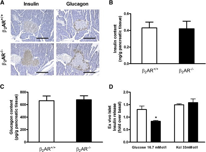
FIG. 2.
Comparison of β2AR+/+ and β2AR −/− pancreatic islets. Immunoistochemical analysis (A) of the islets was carried out on paraffin sections using insulin (left panel) or glucagon (right panel) antibodies. Microphotographs are representative of images obtained from pancreas sections of five 6-month-old β2AR+/+ (upper panel) or β2AR −/− (lower panel) mice. Insulin (B) and glucagon (C) content in isolated islets from β2AR+/+ (n = 10) or β2AR −/− (n = 13) mice. Insulin secretion in response to basal (2.8 mmol/L) or high (16.7 mmol/L) glucose concentration and to KCl (33 mmol/L) was measured in isolated islets from β2AR+/+ (□) and β2AR−/− (■) mice (D). Bars represent means ± SE of data from 10 mice per group. *P < 0.05 vs. β2AR+/+, Bonferroni post hoc test. (See also Supplementary Fig. 1.) (A high-quality digital representation of this figure is available in the online issue.)

