FIG. 4.
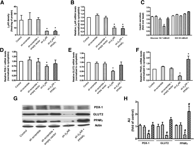
β2AR levels, glucose-stimulated insulin secretion, and gene expression profile in silenced INS-1E β-cells. Treatment with a specific β2AR-shRNA significantly decreased the density (by 73.7% [A]) and mRNA levels (by 59.1% [B]) of β2AR in INS-1E β-cells. β2AR-shRNA inhibited the insulin secretory response to 16.7 mmol/L glucose, which was rescued by the overexpression of PPARγ (C). KCl-induced insulin release (C) was not significantly different among the studied groups. β2AR-shRNA also determined a significant reduction in mRNA level of PDX-1 (D), GLUT2 (E), and PPARγ (F) that was prevented by the overexpression of PPARγ. Bars represent means ± SE from four to five independent experiments in each of which reactions were performed in triplicate ( , control, i.e. untreated INS-1E β-cells;
  , sh-scramble;
  , sh-scramble+empty vector;
  , sh-β2AR;
  , sh-β2AR+PPARγ; *P < 0.05 vs. control, Bonferroni post hoc test; basal is glucose 2.8 mmol/L. Equal amount of proteins from three independent experiments was analyzed by Western blotting and quantified by densitometry (G and H ). *P < 0.05 vs. sh-scramble. AU, arbitrary units. (See also Supplementary Figs. 2–4.)

