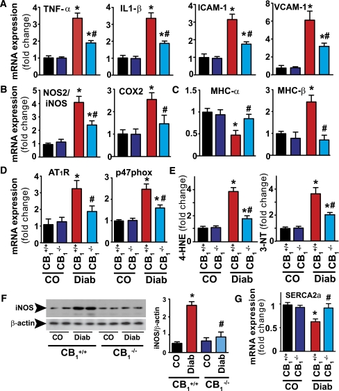
FIG. 2.
Attenuation of diabetes-induced myocardial inflammation, oxidative/nitrative stress, β-MHC isozyme switch, and AT1R expression in CB1−/− mice. A: LV mRNA expressions of inflammatory cytokines and adhesion molecules. B: iNOS and COX2. C: α- and β-MHC. D: AT1R and p47phox NADPH isoform. E: Oxidative/nitrative stress was determined by measuring 4-HNE and 3-NT in the LV myocardial tissues. F: Protein of iNOS in the respective groups. G: SERCA2a. *P < 0.05 vs. WT/CB1+/+ control (CO); #P < 0.05 vs. CB1+/+ diabetes (Diab); n = 8–9/group. (A high-quality color representation of this figure is available in the online issue.)

