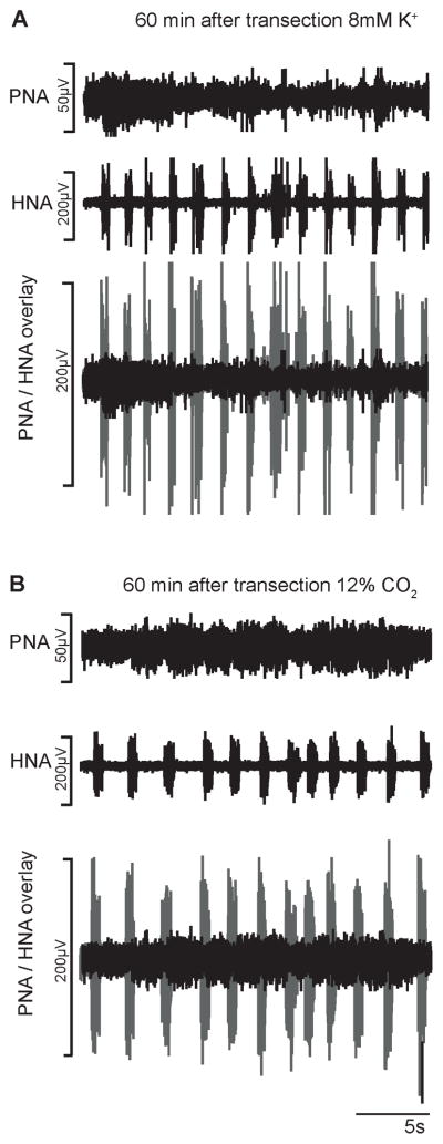
Fig. 3.
Phrenic and hypoglossal nerve recordings (PNA, HNA) 1h after transection. A. Identifiable but sporadic low amplitude PNA bursts after increase of extracellular K+. B. Sporadic PNA bursts during hypercapnia (12% CO2). Comparison of eupneic HNA amplitude and spinal cord generated PNA is illustrated in overlays of PNA (black) and HNA (gray).

