Figure 1.
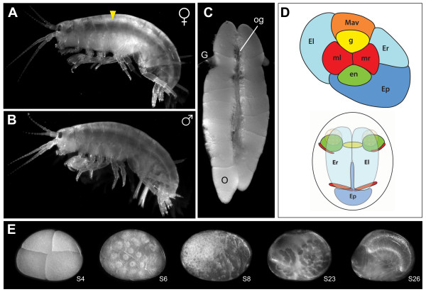
Parhyale hawaiensis and the tissues used to construct a de novo transcriptome. (A) Adult female amphipod, P. hawaiensis. (B) Adult male. (C) Ovaries of adult female. Oocytes and oogonia are visible at various stages of growth. (D) Schematic drawings of the eight cell stage (top), at which all germ layers and the germ line are specified, and the germ band stage (bottom). Both of these signature stages are represented in this transcriptome. (E) A sample of the range of stages of P. hawaiensis embryogenesis represented in this transcriptome; stages as per [55]. Embryos from as early as S1 (one cell stage) and as late as S27 (just before hatching) were sampled; see Additional File 1 for details. G: gonoduct; O: late stage oocyte; og: younger oocytes and oogonia. Anterior is to the left in A, B, and E, and up in C.

