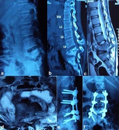
Fig. 1.
Pre-operative lateral (a) X-rays and sagittal reconstructed computed tomography (CT) (b), Midsaggitial T2-W1 (c) and axial (d) magnetic resonance image (MRI) of a 16-year old girl with Pott’s disease at L4–L5 showing reversal of normal lumbar lordosis to kyphosis of 50°. Immediate lateral (e) and anteroposterior (AP) (f) X-ray of the patient after anterior radical debridement and posterior instrumentation using pedicle screw fixation via the transpedicular approach

