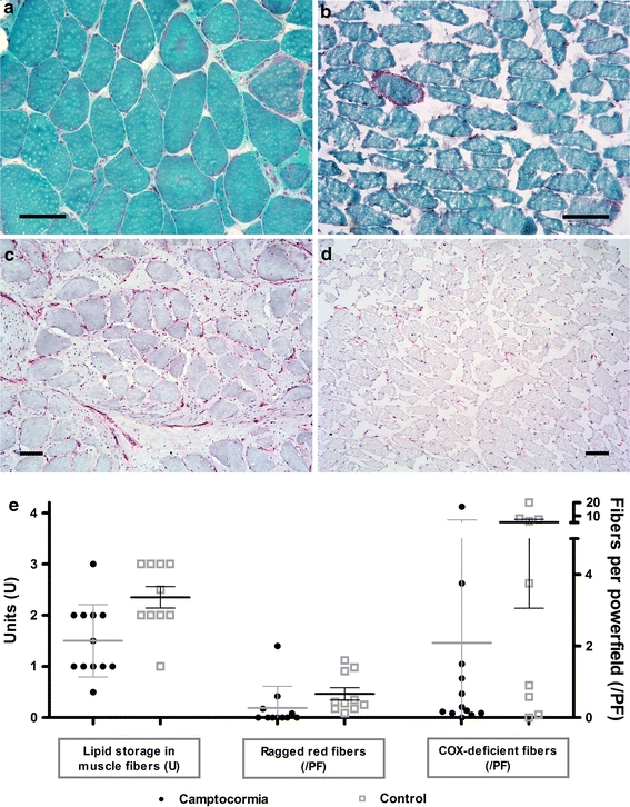
Fig. 4.

Camptocormia shows no evidence for mitochondriopathy or inflammation. Modified Gomori’s trichrome stain sometimes shows a higher content of ragged red and COX-deficient fibers in PD camptocormia biopsy tissues (a, e) than in control limb muscles (see Fig. 1) but the same holds true for the paraspinal control muscles (b, e). No evidence for inflammation was observed (c, d MHC-1 immunohistochemistry). Bars 100 μm
