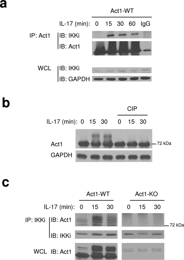
Figure 1. IKKi forms a complex with Act1 upon IL-17 stimulation.
A. Cell lysates from Act1-deficient MEFs infected with retroviral WT Act1 (Act1-WT) untreated or treated with IL-17 (50 ng/ml) for 0, 15, 30 and 60 min were immunoprecipitated with anti-Act1 or IgG, followed by immunoblot analysis with anti-IKKi, anti-Act1 and anti-GAPDH. WCL: whole cell lysates.
B. Lysates from Act1 WT reconstituted MEFs treated with IL-17 (50 ng/ml) for 0, 15 and 30 min were untreated or treated with phosphatase (CIP, 1 h, 37°C)], followed by immunoblot analysis with anti-Act1 and anti-GAPDH.
C. Cell lysates from wild-type and Act1-deficient MEFs untreated or treated with IL-17 (50 ng/ml) for 0, 15 and 30 min were immunoprecipitated with anti-IKKi, followed by immunoblot analysis with anti-Act1 and anti-IKKi. WCL: whole cell lysates.
The data shown in this figure are representation of three independent experiments.

