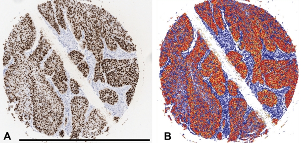
Figure 1.
The picture segmentation demonstrated at cylinder stained for p53 at baseline. Background and counterstaining is blue. Strong positive staining is red (0-100 gray levels) and regarded as true positive. Orange (101-175) and yellow (176-220) were considered negative, mainly constituting cytoplasmic background staining. A-B, bar = 1.0 mm.

