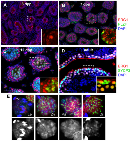
Fig. 1.
BRG1 expression during spermatogenesis. (A-D) BRG1 (red) expression in neonatal testes at specified ages (A–C) and adult testis at 8 weeks (D). Insets show magnified views of the marked areas depicting BRG1 detection in prospermatogonia [round, large nuclei (A), PLZF-positive (green) spermatogonia (B), and SYCP3-positive (green) spermatocytes (C,D)]. (E) Double immunostaining of BRG1 (red) and SYCP3 (green) in nuclei spreads prepared from 8-week-old testis. Spermatocytes were staged based on the appearance of the synaptonemal complex in nuclear spreads (marked by SYCP3, green). Nuclear staining is enhanced by DAPI-only images (bottom panels). Bright and hooked nuclei (arrowhead) are sperm. In all images, nuclei were counterstained with DAPI (blue) unless otherwise specified. Scale bars: 50 μm. Di, diplonema; Le, leptonema; Pa, pachynema; Zy, zygonema.

