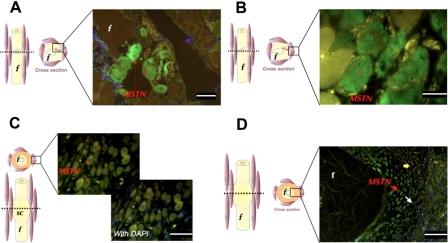
Figure 2.
Immunohistochemistry staining (green fluorescence) of myostatin in transverse section of fracture site. Left-panel figures indicate plane of section and area of interest shown in micrograph. Injured skeletal muscle fibers highly express myostatin at 12 hr (A; scale bar = 25 µm) and 24 hr (B; scale bar = 10 µm) following surgery. Six days postfracture, high myostatin expression (arrow) is restricted to the nuclei of regenerating myotubes (C; scale bar = 25 µm). Four days postfracture, round, mature soft callus chondrocytes (red arrow) and flat proliferating chondrocytes (white arrow) express myostatin (D; scale bar = 50 µm). Sections are counterstained with DAPI, and blue DAPI staining is abundant in the bottom right quadrant of the image (D). f, fibula; MSTN, myostatin; sc, soft callus. Yellow arrows indicate chondroprogenitor cells. Yellow staining represents autofluorescence.

