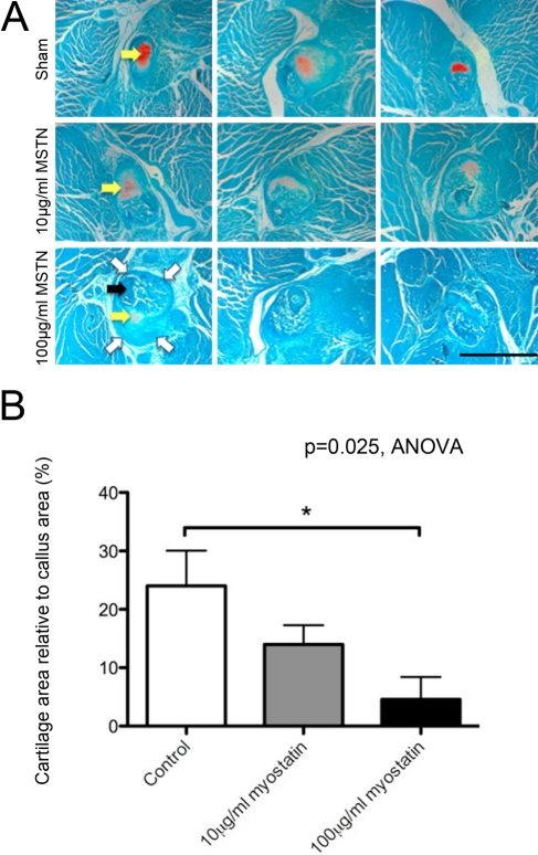
Figure 3.
Endochondral ossification (orange stain, yellow arrows) is apparent at 15 days after fracture and is decreased by local treatment with recombinant myostatin. Light microscopic images of transverse sections in the center of 15-day-old fracture callus of myostatin-treated mice, stained with Safranin O and fast green. Fracture callus outline is indicated by open white arrows and cortical bone of the fractured fibula by the solid black arrow (A; scale bar = 500 µm). Cartilage area relative to total callus area decreased in a dose-dependent manner with myostatin treatment (B; n=5–7 per group). MSTN, myostatin.

