Figure 5.
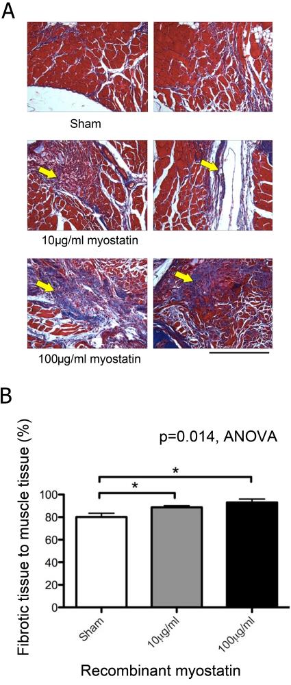
Fibrous tissue formation in local myostatin-treated mice. Histological sections stained with Masson’s trichrome showing a dose-dependent increase in fibrous tissue formation (blue stain, arrow) during muscle repair with myostatin treatment (A; scale bar = 500 µm). Myostatin treatment (10 or 100 µg/ml) significantly increased the relative fraction of fibrous tissue staining blue in trichrome sections relative to vehicle treatment (B; n=5–7 per group).

