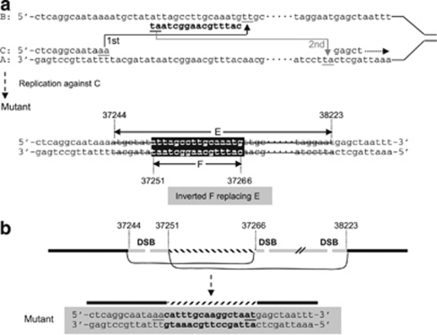
Figure 4.
Illustration of two possible mechanisms underlying the complex rearrangement detected in patient BEA. (a) In the SRS model,42, 43 the newly synthesized leading strand (C) misaligned with the lagging template strand (B) through inverted short repeats (the first step of slippage); having synthesized a short DNA sequence tract, the C strand dissociated from B and re-annealed to its original template strand (A) via short repeats (the second step of slippage); continued DNA synthesis (right-handed dotted arrow). Then replication against the synthesized strand C resulted in the observed complex rearrangement. (b) In the non-homologous end-joining (NHEJ) model, at least two double-strand breaks (DSB) occurred within the sequences deleted. One short internal sequence was re-captured in an inverted orientation during the NHEJ repair. Numbers indicating nucleotide positions are in accordance with GenBank accession EF034088.1. Short repeats are underlined.

