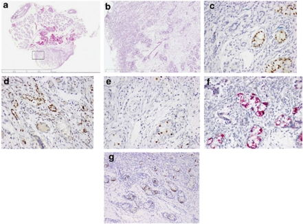
Figure 1.
Representative histological and immunohistochemical findings for the left gonad: (a) total overview of the gonad histology (H&E); (b) higher magnification ( × 2.5 magnification, indicated by square in (a); immunohistochemical detection of (c) SOX9, positive in the Sertoli cells; (d) FOXL2, positive in the granulosa cells; (e) OCT3/4; (f) TSPY; (g) KITL, all positive in the transformed germ cells. All immunohistochemical images are at the magnification of × 100, except G, being × 50.

