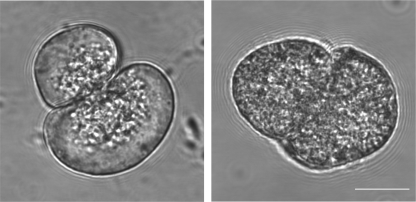
FIGURE 1.
Isolated pancreatic acinar cells show a greatly increased number of zymogen granules in the VAMP8 knock-out mice. Morphological differences in the isolated exocrine pancreas from WT versus VAMP8 knock-out mice have previously been described at low magnification. Here, at high magnification, phase images of isolated single cells (all functional experiments are with pancreatic tissue fragments, but the differences in granule distribution are difficult to see in multicellular fragments) show the clustering of granules in the apical region (where the two cells are touching) of WT cells (left) compared with the distribution of granules across the whole of the cell in VAMP8 knock-out cells (right).

