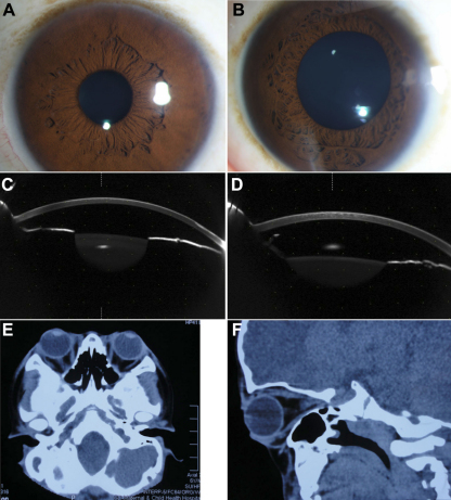
Figure 3.
Examination results of II-3 and III-1. A: Anterior segment photograph of II-3. B: Anterior segment photograph of III-1. C: The anterior segment picture of II-3 by Pentacam. D: The anterior segment picture of III-1 by Pentacam. E, F: Shallow orbits and ocular proptosis of III-1 using computed tomography (CT).

