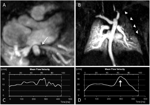
Figure 2.
Premature infant who underwent CMR at 8 weeks corrected gestational age weighing 3.0 kg. The original diagnosis was total anomalous pulmonary venous connections (cardiac type) repaired by the APA. A) Axial image shows severe narrowing of the left upper PV post-operatively (arrow). B) Maximum intensity projection of the magnetic resonance angiography dataset demonstrates delayed filling of the left lung, particularly of the upper lobe (arrowheads). The left upper PV is not shown on this first pass angiographic acquisition, but filled during the second acquisition 12 seconds later. C) Time-velocity flow curve in the left lower PV reveals loss of phasic flow at a relatively low velocity (proximal to the narrowing). D) Normal post-operative flow pattern in the common right PV in the same patient, with two distinct peaks, the greater one during diastole (arrow).. Only 27% of pulmonary blood flow went to the left lung. Note the different velocity scales for the left (C) and right (D) PV flows.

