
Figure 3.
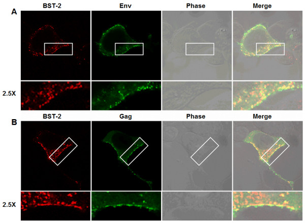
MMTV Gag co-localizes with BST-2. 293T cells transiently transfected with BST-2 and MMTV proviral constructs were permeabilized, fixed, stained, and analyzed with confocal microscopy for BST-2 and (A) MMTV Env or (B) MMTV Gag localization. Green is MMTV Env or Gag, red is BST-2, orange is merge. Phase contrast shows cell morphology. Areas of BST-2/Env or BST-2/Gag co-localization at the cells surface are highlighted in white rectangular boxes and magnified by 2.5X. Experiments were repeated at least three times with similar results.