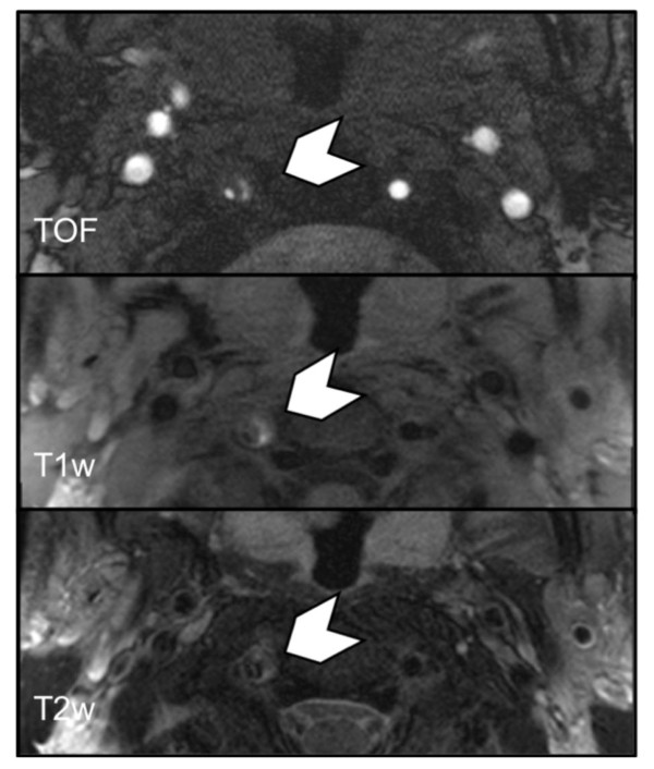
Figure 4.
Multi sequence CMR showing VWH (chevron) of the right VA in the V2 segment with mixed signal intensities in a 34-year-old patient with sCAD. The hematoma presents with imaging features of acute, early subacute and late subacute hemorrhage. The VWH was classified according to the oldest hemorrhage type present (i.e. late subacute). CMR interval from onset of symptoms to scan was 9 days.

