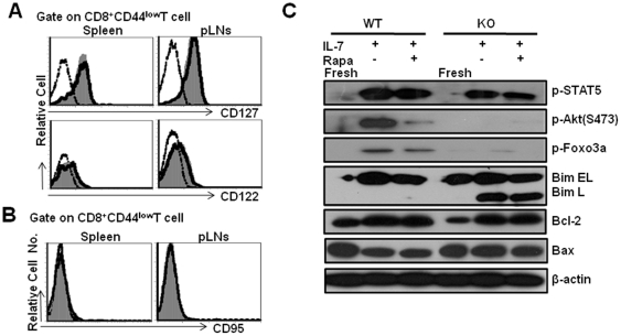
Figure 5. Decreased Akt-FoxO1/FoxO3a phosphorylation of Tsc1 KO naïve CD8+T cells in response to IL-7.
The expression of CD122, CD127 and CD95 on naïve CD8+T cells was determined by gating CD8+CD44low cells. Comparable surface CD122, CD127 (A) and CD95 (B) expression in Tsc1 KO naïve CD8+ T cells. The dash-dot line represents staining with an isotype control antibody. The histograms (gray) represent WT whereas the open histograms with solid line show Tsc1 KO staining pattern. Cells are gated with CD8+CD44low population. Representative data are shown from one of two separate experiments, with three mice in each group. (C) Western blot analysis of mTORC2-Akt-FoxO1/FoxO3a-Bim axis in naïve CD8+ T cells after stimulation with IL-7. Sorted naïve CD8+T cells were cultured with IL-7 in the presence of Rapa or not for 24 hrs. The freshly isolated WT or Tsc1 KO naïve CD8+T cells were used as a control. One representative is shown from two or three separate experiments.

