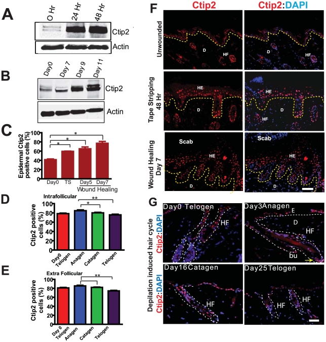
Figure 1. Expression of Ctip2 in skin of adult mice following tape stripping (TS), during full thickness wound healing and in hair cycling.
Immunoblot analyses of Ctip2 protein expression in wild type mice skin 24 and 48 hrs post tape stripping (A), and in wound biopsies obtained at Days 7, 9 and 11 after wounding (B). β-actin is used as control. (C) Bar chart showing an increase in the percentage of Ctip2 positive cells 48 hours post TS and on days 5 & 7 post wounding (*p<0.05). Bar chart showing, percentage of (D) intrafollicular and (E) extrafollicular Ctip2 expressing cells in depilation induced hair cycling in adult mouse skin. Significant (* P<0.05; **P<0.005) increase in Ctip2 expression was observed in induced anagen compared to telogen and catagen stages (D and E). (F) An overview of Ctip2 localization by immunofluorescence (IF) in the adult unwounded mice skin, 48 hr after mechanical injury and at day 7 post wounding using anti-Ctip2 antibody. (G) Overview of Ctip2 localization by IF in depilation induced hair cycle in mice skin. HE-hyperproliferative epithelium; HF-hair follicle; E-epidermis; D-dermis; Bu-bulge; DP-dermal papilla; bu-bulge. Yellow dotted lines separates the epidermis from dermis (F) and white dotted lines outlines the HF (G). Scale bars: 50 µm (F) and 25 µm (G).

