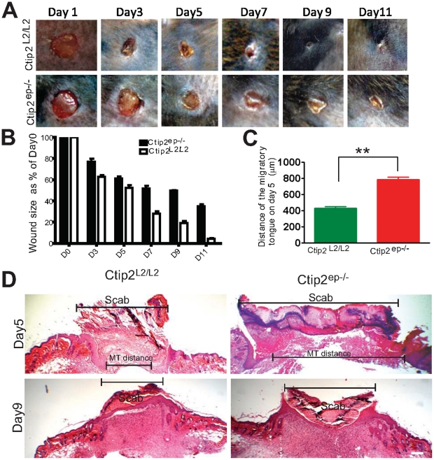
Figure 2. Impaired wound healing in skin of adult Ctip2ep−/− mice.
(A) Macroscopic images of time course of healing of 5 mm full-thickness excisional wounds. Note, that in Ctip2L2/L2 mice at day 7, 9 and 11 post-wounding, the wound area was significantly reduced compared to the Ctip2ep−/− mouse. (B) At indicated time points, the diameter of the wound was measured using photography and digital analyses. Data from three independent experiments (9 Ctip2L2/L2 mice and 9 Ctip2ep−/− mice were used for this study) were pooled. The difference in the rate of wound closure was statistically significant between two groups (P<0.001). (C) Graph showing increased migratory tongue distances between the two migratory epithelial tongue on the wound bed from Ctip2L2/L2 and Ctip2ep−/− mice. (*p<0.05). (D) Hematoxylin and Eosin (H & E) stained images of the day 5 and 9 post wounding samples. The black broken brackets in day 5 and day 9 show the outline of the wound area and the migratory tongue distance (day 5). Hyperproliferative epithelium; E- Epidermis; D-Dermis. Scale bar: 200 µm (D).

