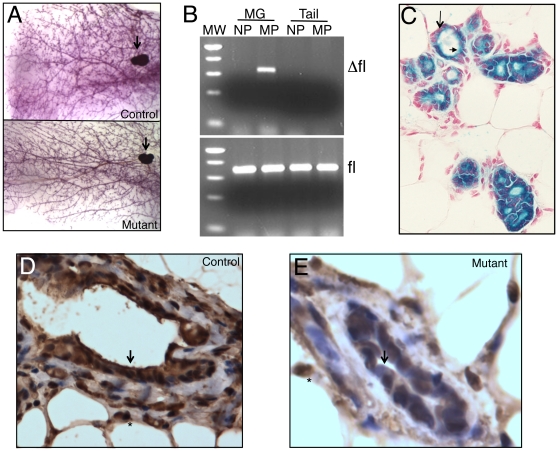
Figure 2. Lineage-specific deletion of Brg1 in the mammary gland.
A. Whole-mount preparation of control (top) and mutant (bottom) mammary glands from multiparous females. Asterisks, lymph nodes. B. Ethidium bromide-stained gels showing Brg1 Δfl (top) and fl (bottom) PCR products. MW, molecular-weight standard (500-, 400-, 300-, 200-, and 75-bp fragments are visible); MG, mammary gland; NP, nulliparous; MP, multiparous. C. Mammary gland section from multiparous mouse carrying the Wap-Cre transgene on a R26R background. Cre activity, visualized as blue X-Gal staining, is restricted to luminal cells (arrowhead). Basal/myoepithelial cells (arrow) are negative and appear pink because of nuclear fast red counterstain. D, E. IHC staining of BRG1 showing strong staining in nuclei throughout the mammary gland in controls (D) but absent in luminal cells of mutant mice (E). Arrows, luminal cells; asterisks, adipocyte nuclei. 400× magnification.

