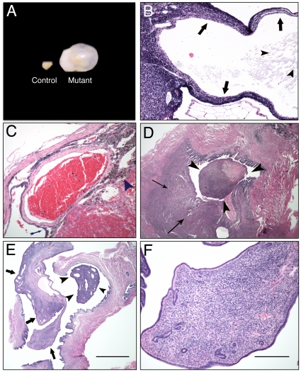
Figure 4. Histopathology of ovarian cysts and uterine tumors from Brg1Wap-Cre mice.
A. Control ovary (left) and cystic ovary from a conditional Brg1Wap-Cre mutant (right). B. Section of an ovarian cyst stained with H&E at 40× magnification. The thin wall of this multilocular structure is comprised of an inner layer of spindle-shaped cells surrounded by multiple layers of round cells with uniformly sized, round, basophilic nuclei with scant cytoplasm (arrows). This cyst is filled with a lightly eosinophilic proteinaceous fluid (arrowheads). C. Hemorrhagic ovarian cyst section stained with H&E at 40× magnification. The thin arrow points to a wall of the cyst, and the arrowhead points to a region of hemosiderin-laden macrophages at the edge of the hemorrhage. D. Histiocytic sarcoma section stained with H&E at 20× magnification. A pleomorphic population of cells with basophilic round to ovoid nuclei and cytoplasm varying from scant to abundant and foamy are seen infiltrating the myometrium (thin arrows) and forming a polyp (arrowheads) projecting into the uterine lumen E. Endometrial stromal polyp section stained with H&E at 20× magnification. The polyp (arrows) includes a pedunculated mass (arrowheads) that projects into the uterine lumen. F. Endometrial stromal polyp section stained with H&E stained at 200× magnification. The polyp stroma is comprised of spindle cells with variable amounts of cytoplasm and ovoid nuclei; the structure also includes small blood vessels and small endometrial glands and is covered by a single layered cuboidal to columnar epithelium with basally located round nuclei.

